Skip to content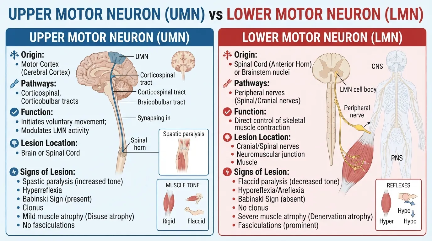
UMN vs LMN refers to the difference between upper motor neurons and lower motor neurons, which work together to control voluntary movement.
UMNs carry motor signals from the brain through descending pathways, while LMNs carry signals directly to skeletal muscles.
UMN lesions commonly cause spasticity and increased reflexes, while LMN lesions commonly cause weakness, reduced reflexes, muscle atrophy, and fasciculations.
Key Takeaways
- UMN stands for upper motor neuron, while LMN stands for lower motor neuron.
- UMNs help send movement commands from the brain to the spinal cord or brainstem.
- LMNs directly connect the nervous system to skeletal muscles.
- UMN lesions commonly cause spasticity, hyperreflexia, and a Babinski response.
- LMN lesions commonly cause flaccidity, hyporeflexia, muscle atrophy, and fasciculations.
What Are UMNs and LMNs?
The nervous system uses several pathways to control movement. Two important parts of this system are upper motor neurons (UMNs) and lower motor neurons (LMNs).
They work as a chain. UMNs carry movement commands from higher areas of the central nervous system toward the spinal cord or brainstem. LMNs then carry those signals to skeletal muscles, where they help produce contraction.
A simple way to picture the pathway is:
Brain → UMN pathway → LMN → muscle
This distinction is important during a neurological examination because damage to each part can produce a different pattern of weakness, reflexes, and muscle tone.
What Does an Upper Motor Neuron Do?
Upper motor neurons are found within the central nervous system. Major descending motor pathways include the corticospinal and corticobulbar tracts.
These pathways help control voluntary movement. UMN signals travel from the brain toward motor neurons in the brainstem or spinal cord.
UMNs do not directly leave the central nervous system to connect with skeletal muscle. Instead, they influence LMNs, which provide the final motor connection to the muscle.
What Does a Lower Motor Neuron Do?
Lower motor neurons provide the direct connection between the nervous system and skeletal muscle.
Their cell bodies are found in areas such as the anterior horn of the spinal cord and certain cranial nerve nuclei. Their axons travel outward and directly innervate muscles.
This is why damage to an LMN can directly affect the muscle it supplies.
UMN vs LMN: Key Differences
The easiest way to understand UMN vs LMN is to compare their location and the signs that appear when each is damaged.
| Feature | UMN | LMN |
| Full name | Upper motor neuron | Lower motor neuron |
| Main location | Brain and descending CNS pathways | Anterior horn, cranial nerve nuclei, peripheral motor pathways |
| Main role | Carries and controls motor commands | Directly activates skeletal muscle |
| Muscle tone after established lesion | Usually increased | Usually decreased |
| Deep tendon reflexes | Usually increased | Usually decreased |
| Weakness | Common | Common |
| Muscle atrophy | Usually limited in isolated lesions | Common and often prominent |
| Fasciculations | Usually absent in isolated UMN lesions | Can occur |
| Babinski response | May be present | Not a typical LMN sign |
| Typical motor pattern | Spastic weakness | Flaccid weakness |
These are general clinical patterns. The exact findings depend on the location, severity, timing, and cause of the neurological lesion.
UMN Lesion Signs
An UMN lesion affects the descending motor pathways above the lower motor neuron. The resulting pattern is often called an upper motor neuron syndrome.
Common findings include weakness, spasticity, hyperreflexia, and clonus. A Babinski response may also occur.
Increased Reflexes and Spasticity
One of the classic differences in an UMN lesion is an increase in muscle tone and deep tendon reflexes.
Hyperreflexia means that reflexes are more active than expected. Spasticity refers to increased muscle resistance associated with certain movements.
This happens because descending motor pathways normally help regulate spinal reflex activity. Damage to those pathways can reduce that control and lead to exaggerated reflex responses.
Clonus, which consists of rhythmic involuntary muscle contractions after a sudden stretch, can also occur with UMN lesions.
Babinski Sign and Weakness
A Babinski response is another important UMN finding. In adults, an abnormal response involves extension of the great toe after stimulation of the sole of the foot. It can indicate corticospinal tract dysfunction.
UMN weakness often affects groups of muscles rather than a single muscle supplied by one peripheral nerve. Fine motor control can also be impaired, particularly in the hands.
An important exception is the early phase after some acute spinal cord injuries. Spinal shock can temporarily cause low muscle tone and absent reflexes before typical UMN features develop.
LMN Lesion Signs
An LMN lesion affects the motor pathway that directly connects with muscle. Common findings include weakness, decreased muscle tone, reduced reflexes, muscle atrophy, and sometimes fasciculations.
Muscle Atrophy and Fasciculations
Because LMNs directly supply skeletal muscles, damage can lead to loss of muscle bulk over time.
Muscle atrophy means the muscle becomes smaller. Fasciculations are small, involuntary muscle twitches that may be visible beneath the skin. They can occur with LMN disorders, although fasciculations can also occur in some people without a serious neurological disease.
The combination of weakness, atrophy, and fasciculations can therefore provide useful clues during a neurological examination.
Reduced Reflexes and Flaccidity
LMN damage can interrupt the reflex arc. As a result, deep tendon reflexes may become reduced or absent, a finding called hyporeflexia or areflexia.
Muscle tone also tends to decrease, producing flaccid weakness. Merck Manual notes that lower motor neuron dysfunction can produce hyporeflexia, decreased muscle tone, fasciculations, and muscle atrophy over time.
Why Are UMN and LMN Signs Different?
The difference comes from where the damage occurs in the motor pathway.
UMNs are part of the descending system that helps control and regulate spinal motor circuits. When this control is disrupted, reflex activity can become exaggerated, producing findings such as hyperreflexia and spasticity.
LMNs are part of the final pathway to the muscle. If they are damaged, the muscle loses direct neural input. This can produce reduced reflexes, low tone, weakness, atrophy, and fasciculations.
In simple terms:
UMN problem → too little descending control of reflexes
LMN problem → loss of the direct motor connection to muscle
This is a useful learning framework, but neurological examination findings should always be interpreted as a complete pattern rather than by one sign alone.
Common Conditions Associated With UMN and LMN Lesions
Many different disorders can affect motor pathways.
Conditions involving the brain or spinal cord can produce UMN findings. Examples include certain strokes, spinal cord disorders, multiple sclerosis, and other diseases affecting descending motor pathways.
LMN findings can occur when disease or injury affects the anterior horn cells, nerve roots, peripheral motor nerves, or related structures. Peripheral neuropathies are one example of disorders that may produce LMN-type findings.
Some neurological diseases can involve both UMNs and LMNs. Amyotrophic lateral sclerosis (ALS) is a well-known example, with clinical findings that can include both upper and lower motor neuron features.
Therefore, seeing one sign alone does not automatically identify a particular disease.
How Clinicians Distinguish UMN From LMN Signs
During a neurological examination, clinicians look at several features together.
They may assess:
- Muscle strength : Is there weakness, and which muscles or muscle groups are affected?
- Muscle tone : Is tone increased or decreased?
- Deep tendon reflexes : Are reflexes normal, exaggerated, or reduced?
- Plantar response : Is there an abnormal Babinski response?
- Muscle bulk : Is there visible atrophy?
- Fasciculations : Are there involuntary muscle twitches?
- Distribution : Does the weakness fit a brain, spinal cord, nerve-root, or peripheral-nerve pattern?
Merck Manual emphasizes that the pattern of weakness, reflexes, tone, and associated neurological findings helps clinicians localize where the problem may be occurring.
UMN vs LMN: Easy Way to Remember the Difference
A simple memory aid is:
UMN = Upper + Upregulated reflexes
Think of UMN lesions as commonly producing:
- Increased tone
- Increased reflexes
- Spasticity
- Babinski response
For LMN, think:
LMN = Low tone + Muscle wasting
Common LMN findings include:
- Decreased tone
- Decreased reflexes
- Muscle atrophy
- Fasciculations
This memory aid is useful for studying, but it should not replace the full neurological examination.
FAQs:
Is UMN spastic or flaccid?
Established UMN lesions are generally associated with spasticity and increased muscle tone. They commonly produce hyperreflexia as well. However, acute severe spinal cord injury can initially cause spinal shock, with temporary flaccidity and reduced reflexes before typical UMN signs emerge.
Is LMN hyperreflexic or hyporeflexic?
LMN lesions are typically hyporeflexic, meaning the affected deep tendon reflexes are reduced. Severe disruption of the reflex arc can result in absent reflexes. LMN lesions can also cause decreased muscle tone, weakness, atrophy, and fasciculations.
Does UMN cause muscle atrophy?
An isolated UMN lesion does not usually cause the marked muscle atrophy associated with LMN damage. Some muscle wasting can develop from reduced use over time, but prominent atrophy and fasciculations are more characteristic of LMN involvement.
Can a disease affect both UMNs and LMNs?
Yes. Some neurological disorders affect both parts of the motor system. ALS, for example, can produce a mixture of UMN and LMN signs.
What is the main difference between UMN and LMN?
The main difference is their position and role in the motor pathway. UMNs carry descending motor commands within the central nervous system, while LMNs directly connect the nervous system with skeletal muscles.
Their lesions therefore produce different patterns of reflexes, muscle tone, atrophy, and weakness.
Conclusion:
Understanding UMN vs LMN makes neurological examination findings much easier to interpret.
UMNs are part of the descending motor pathways that control and regulate movement, while LMNs provide the final direct connection to skeletal muscles.
In general, UMN lesions are associated with spasticity, hyperreflexia, and abnormal plantar responses, while LMN lesions are associated with flaccidity, hyporeflexia, atrophy, and fasciculations.
The distinction is useful for understanding neurological disorders and locating where a motor pathway may be affected.
Actual diagnosis, however, requires a complete clinical evaluation and, when appropriate, additional testing.

I am Caleb Anderson, a Christian writer passionate about exploring deep biblical meanings and spiritual truths.
On Biblicaltribez.com, I share faith based insights to help readers grow closer to God through Scripture.
My writing focuses on making the Bible easy to understand, meaningful, and relevant to everyday life.