Skip to content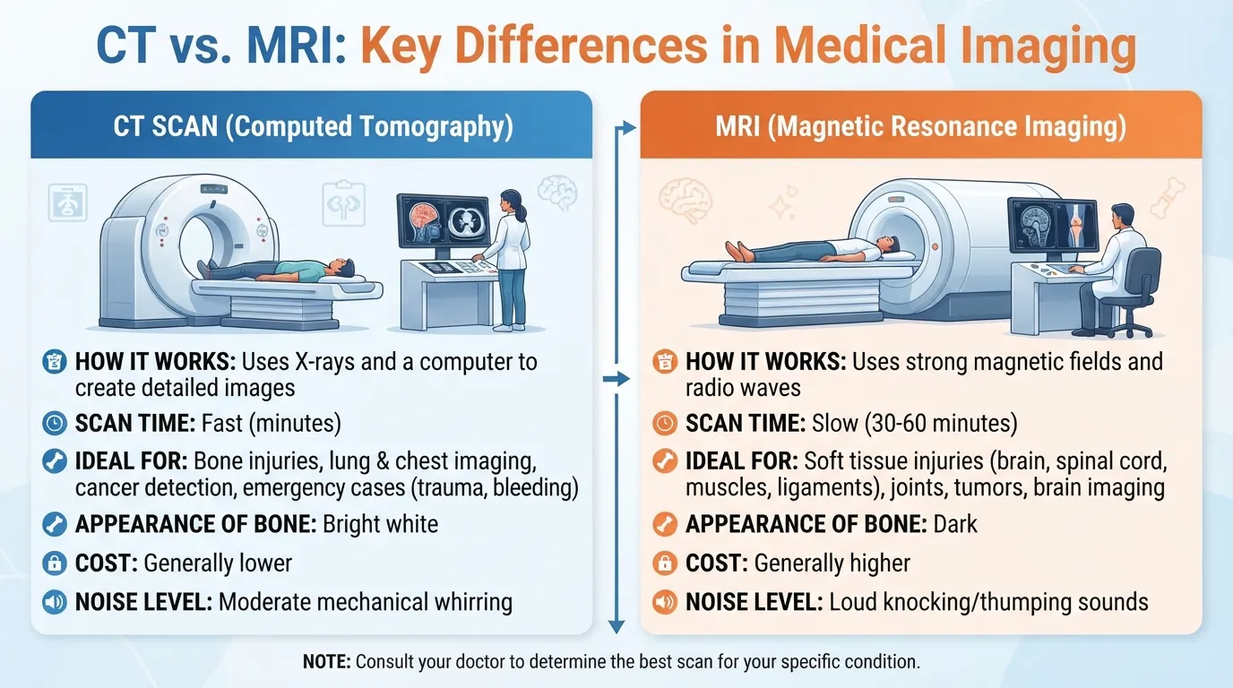
CT vs MRI: A CT scan uses X-rays to create fast, detailed images of bones, organs, blood vessels, and other structures.
MRI uses strong magnetic fields and radio waves and provides greater soft-tissue detail without ionizing radiation.
Doctors choose between them based on the body area, suspected condition, urgency, and patient-specific safety factors.
Key Takeaways
- CT uses X-rays, while MRI uses magnetic fields and radio waves.
- CT is usually faster and is commonly used when doctors need quick answers.
- MRI generally provides more detailed images of many soft tissues.
- CT involves ionizing radiation; MRI does not.
- MRI safety requires careful screening for certain implants and metal objects.
- Neither test is always better. The right choice depends on the medical question.
What Is the Difference Between CT and MRI?
CT and MRI are both medical imaging tests. They help doctors see structures inside the body without surgery, but they create those images in different ways.
CT, or computed tomography, uses multiple X-ray images and a computer to create cross-sectional pictures, often called slices. These images can also be reconstructed into different planes or 3D views. CT can show bones, organs, blood vessels, and other tissues.
MRI, or magnetic resonance imaging, uses a strong magnetic field and radio waves to create detailed images. MRI is especially useful when doctors need a closer look at soft tissues such as the brain, spinal cord, muscles, ligaments, and tendons.
How CT Works
During a CT scan, an X-ray source and detectors rotate around the body. A computer processes the information into detailed cross-sectional images.
CT is fast and relatively less affected by patient movement than MRI. This makes it particularly useful in emergency settings.
How MRI Works
During an MRI, you lie on a table that moves into a large scanner. The machine uses magnetic fields and radio waves to produce images.
MRI does not use ionizing radiation. Depending on the body area and purpose of the examination, an MRI can take roughly 20 to 90 minutes.
CT vs MRI: Key Differences
| Feature | CT | MRI |
| Full name | Computed tomography | Magnetic resonance imaging |
| Main technology | X-rays | Magnetic fields and radio waves |
| Ionizing radiation | Yes | No |
| Soft-tissue detail | Good | Often better |
| Bone imaging | Excellent | Less commonly preferred |
| Speed | Usually faster | Usually slower |
| Movement sensitivity | Less sensitive | More sensitive |
| Metal/implant concerns | Usually fewer | Important safety consideration |
| Common emergency use | Very common | More limited by scan time and availability |
The important point is that these tests are complementary, not simply competing versions of the same test. The best choice depends on what doctors need to see and how quickly they need the information.
When Is a CT Scan Usually Used?
CT is often selected when speed and detailed cross-sectional imaging are important.
Emergency and Trauma
CT is commonly used after serious injuries because it can quickly identify internal bleeding, fractures, and other injuries. It is also frequently used when doctors need rapid information about the head, chest, abdomen, or pelvis.
For example, someone who arrives at an emergency department after a major accident may receive a CT because doctors may need answers quickly.
Bones, Lungs, and Blood Vessels
CT provides excellent information about bones and can also show lungs and blood vessels in considerable detail.
Specialized CT examinations, such as CT angiography, can be used to examine blood vessels. CT is also widely used for chest and abdominal imaging.
When Is an MRI Usually Used?
MRI is particularly useful when detailed soft-tissue information is needed.
Brain, Spine, and Nerves
MRI can provide detailed images of the brain, spinal cord, and other nervous-system structures. It is often useful for evaluating tumors, certain strokes, and other neurological conditions.
CT may still be preferred in an emergency because it is faster. For example, when doctors suspect bleeding after a head injury, CT can provide rapid information. MRI may be used later when more detailed information about brain tissue is needed.
Muscles, Joints, and Other Soft Tissues
MRI is commonly used to examine muscles, tendons, ligaments, joints, and other soft tissues.
For example, an MRI may help evaluate a knee ligament injury or problems involving the spine. MedlinePlus notes that MRI is often used for conditions affecting soft tissues, including the brain, spinal cord, muscles, ligaments, tendons, and joints.
CT vs MRI for the Brain
Both CT and MRI can be used to examine the brain, but they are useful for different reasons.
CT may be preferred when:
- A rapid scan is needed.
- A head injury is involved.
- Doctors need to check for acute bleeding.
- A skull injury is suspected.
- An emergency department needs fast imaging.
MRI may be preferred when:
- Detailed brain-tissue images are needed.
- A tumor or certain neurological conditions are being evaluated.
- More sensitive imaging is needed for some types of stroke.
- Doctors need detailed information about the brain’s structure.
Mayo Clinic explains that CT is often the first brain-imaging test when speed is critical, while MRI can provide more detailed images of brain tissue.
Which Is Faster: CT or MRI?
CT is generally faster than MRI.
A CT examination can often be completed quickly, which is one reason it is valuable in emergency care. MRI examinations usually take longer because multiple image sequences may be needed, and the patient must remain still. The FDA notes that a typical MRI scan can last about 20 to 90 minutes, depending on the body area being examined.
The exact time varies by the type of examination, the body part being scanned, whether contrast is required, and the facility.
CT vs MRI: Radiation and Safety
One of the biggest differences between CT and MRI is radiation.
CT Radiation
CT uses X-rays, which are a form of ionizing radiation. The radiation exposure from a medically appropriate CT scan carries a small potential long-term risk, and the amount depends on the examination. The FDA recommends using CT when it is clinically justified and minimizing unnecessary radiation exposure.
This does not mean a necessary CT should automatically be avoided. Doctors consider the expected medical benefit when deciding whether the examination is appropriate.
MRI Safety and Metal Implants
MRI does not use ionizing radiation, but its powerful magnetic field creates different safety concerns.
Certain metal objects and medical devices can be attracted by the magnetic field, heat up, malfunction, or interfere with the images. Patients should tell the imaging team about implants, pacemakers, metal fragments, and other devices before an MRI. The device’s specific MRI safety status matters.
What About Contrast Dye?
Both CT and MRI can sometimes use contrast material, but the substances are different.
CT commonly uses iodinated contrast material, while MRI may use gadolinium-based contrast agents. Contrast can make certain tissues, blood vessels, or abnormalities easier to see.
Contrast is not required for every scan.
Tell your healthcare provider about kidney problems, previous reactions to contrast, allergies, pregnancy, or other relevant medical conditions before an examination. The imaging team can determine whether contrast is appropriate.
CT vs MRI During Pregnancy
Pregnancy is an important factor when selecting medical imaging.
MRI does not use ionizing radiation, and current radiology guidance states that there are no proven risks to the fetus from MRI itself. CT can also be performed during pregnancy when medically necessary, but because it uses X-rays, doctors consider whether another imaging method can answer the medical question.
If you are pregnant or might be pregnant, tell your healthcare provider before imaging. A necessary examination should not be delayed simply because you are concerned about radiation; the medical team can weigh the risks and benefits for your particular situation.
Is CT or MRI Better?
There is no universal answer to whether CT or MRI is better.
CT may be more useful when speed is critical, when bone needs to be examined, or when certain emergency conditions need rapid evaluation.
MRI may be more useful when detailed soft-tissue information is the main goal and there is enough time for a longer examination.
The American College of Radiology’s Appropriateness Criteria show that imaging choices depend on the specific clinical situation rather than applying one test to every patient.
Your doctor or radiologist considers your symptoms, the body area being examined, the suspected condition, previous imaging, urgency, implants, pregnancy status, and other safety factors.
FAQs:
Is MRI safer than CT?
MRI does not expose patients to ionizing radiation, while CT uses X-rays. However, MRI has its own safety considerations because of its powerful magnetic field, particularly for some implants and metal objects. Therefore, “safer” depends on the patient and the clinical situation.
Which is better for soft tissue?
MRI generally provides better soft-tissue contrast than CT. This makes MRI especially useful for many examinations involving the brain, spinal cord, muscles, ligaments, and tendons. CT can still provide valuable soft-tissue information and may be preferred when speed is important.
Which is better for bones?
CT is generally preferred when detailed bone imaging is needed. It can provide highly detailed cross-sectional images of bones and is commonly used for fractures and other bone-related problems.
Can MRI replace CT?
Not always. MRI and CT answer different medical questions. CT may be necessary when rapid imaging is needed or when certain bones, lungs, bleeding, or other structures need evaluation. MRI may provide information that CT cannot show as clearly, especially for many soft tissues.
Conclusion:
CT and MRI are both important diagnostic imaging tools, but they work differently and have different strengths. CT uses X-rays and is often chosen when doctors need fast imaging, particularly in emergencies. MRI uses magnetic fields and radio waves and provides excellent soft-tissue detail without ionizing radiation.
The right test depends on the medical question, body area, urgency, safety considerations, and information the healthcare team needs. If your doctor has recommended a CT or MRI, ask what they are looking for and why that particular test is appropriate for your situation.

I am Samuel Brooks, a Christian author who enjoys breaking down complex Bible concepts into simple truths. At Biblicaltribez.com, I write content that inspires faith, hope, and spiritual growth. I aim to help readers connect Scripture with real life challenges in a meaningful way.